Spatial organization of malignant and immune compartments in HNSCC
SPATIAL TRANSCRIPTOMICS · PORTFOLIO PROJECT
How are malignant and immune compartments spatially organized across two HNSCC tissue sections?
This project combines sample-level Seurat analysis, reciprocal PCA integration and SpaCET deconvolution to describe spatial transcriptional domains and sample-relative immune niches in two 10x Genomics Visium sections.
Project at a glance
2 tissue sections
HNSCC Visium samples 17B5776 and 19H1257, analyzed independently before joint comparison.
4 analysis stages
Quality control, individual clustering, RPCA integration and spatial deconvolution.
Sample-relative niches
Immune-high and Immune-low spots are defined from quartiles calculated separately within each section.
Reproducible outputs
Saved objects, marker tables, SpaCET summaries, spatial figures and a scientific report support the portfolio narrative.
Computational workflow
01
Import and QC
Load Space Ranger counts and tissue coordinates; inspect spot-level transcript, feature and mitochondrial metrics.
02
Normalize and cluster
Apply SCTransform, PCA, graph-based clustering and marker-guided spatial-domain assessment within each sample.
03
Integrate samples
Use a joint representation and RPCA integration to describe shared and sample-associated transcriptional structure.
04
Resolve compartments
Use SpaCET to estimate malignant, stromal and immune fractions, then map sample-relative immune niches.
Selected findings
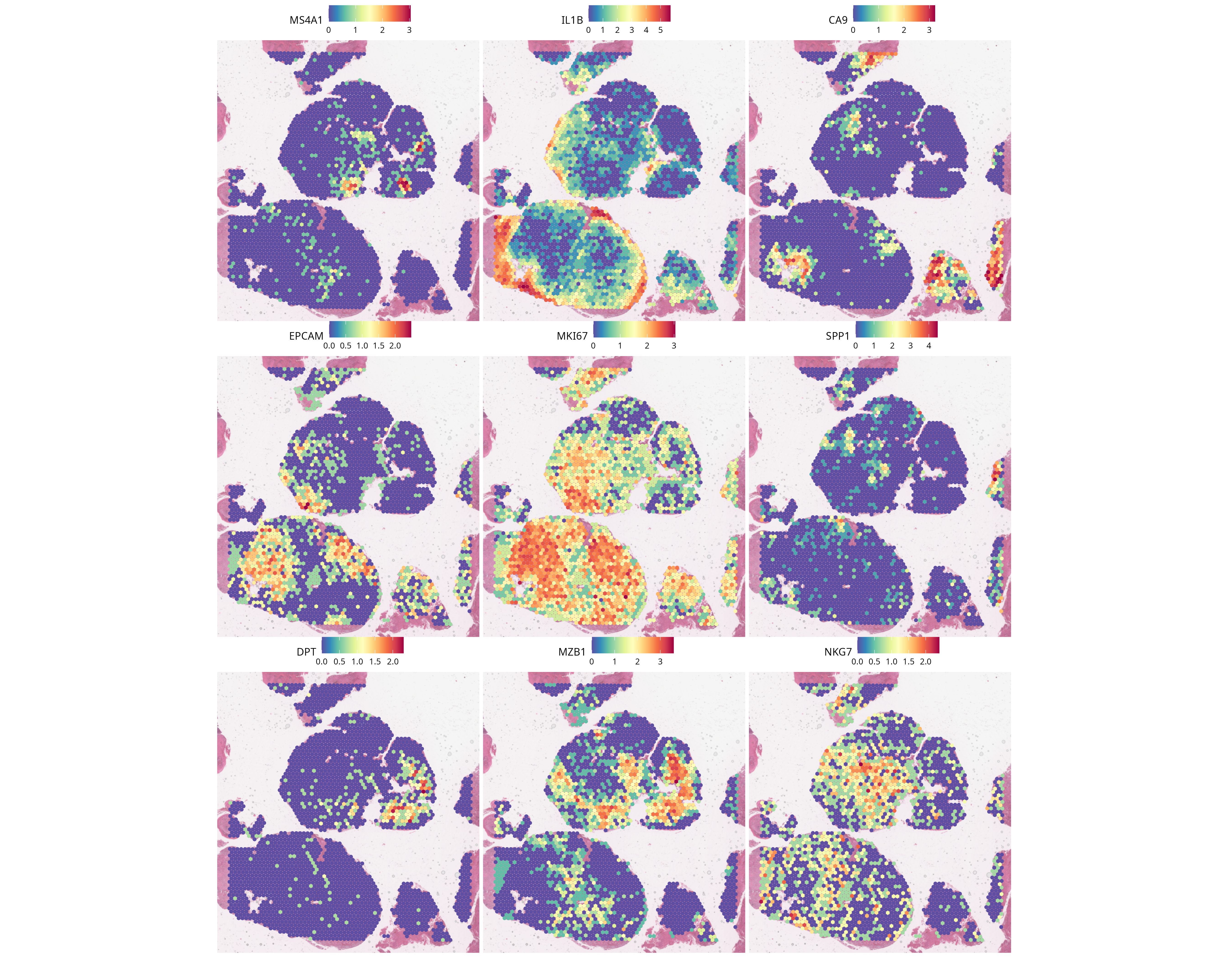
Spatial transcriptional heterogeneity
Observation. Sample 17B5776 contains spatially organized marker programs associated with immune, epithelial, stromal, hypoxic, inflammatory, interferon-responsive and proliferative domains.
Interpretation. These are provisional mixed-spot domain annotations, not pure cell types or formally defined malignant states.
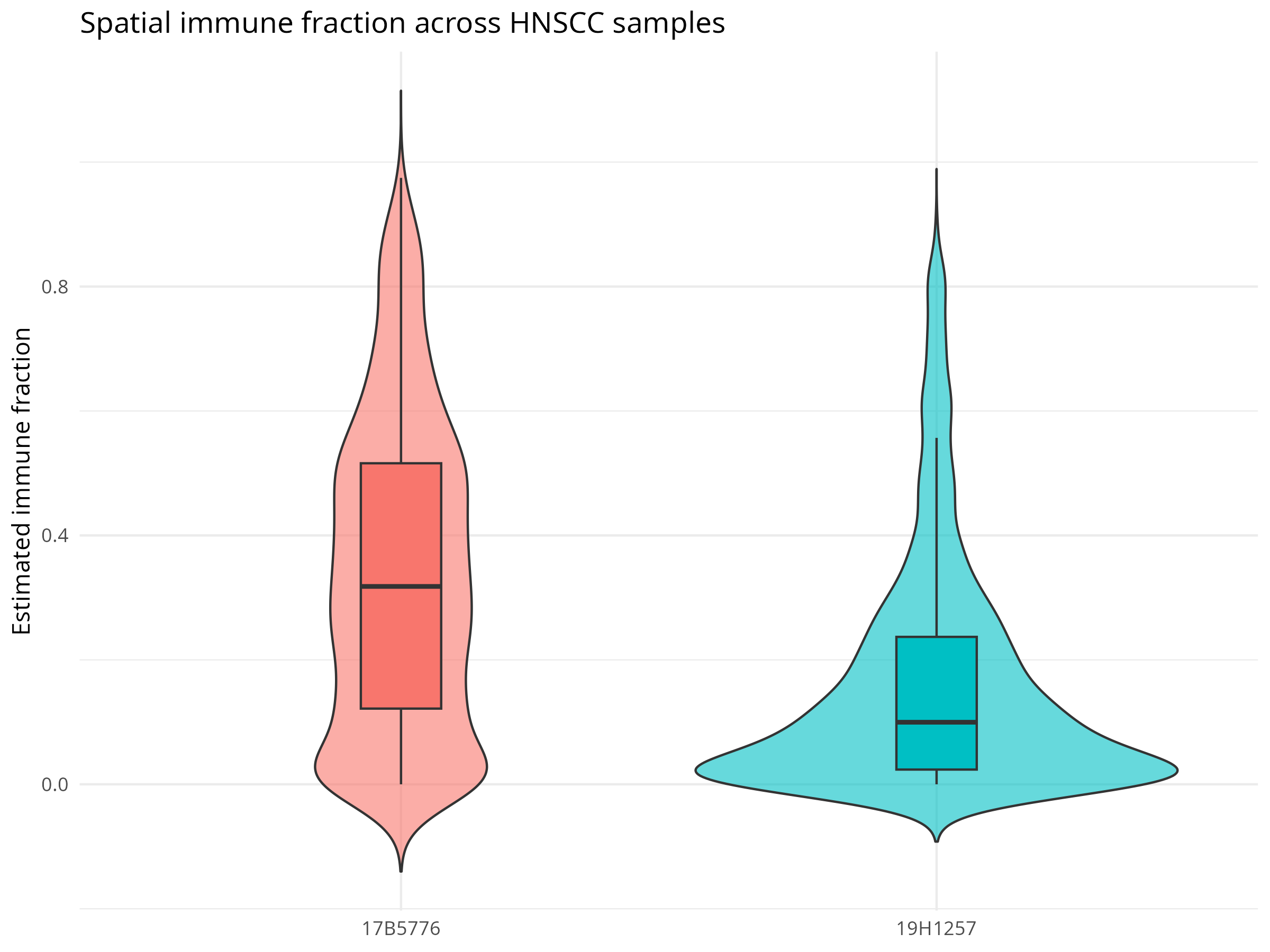
Contrasting immune distributions
Observation. The saved SpaCET summary reports a higher median derived immune fraction in 17B5776 than in 19H1257.
Interpretation. This is a descriptive contrast between two sampled sections and cannot be generalized to an HNSCC population.
The current workflow characterizes malignant fractions and immune niches, but it does not formally identify distinct malignant transcriptional states. Estimated malignant fractions and epithelial marker patterns should not be presented as a completed malignant-state analysis.
Explore the project
Results
Spatial figures, compartment summaries, sample comparisons and downloadable result tables.
Methods
Dataset, QC, normalization, clustering, integration, SpaCET and niche definitions.
Full scientific report
Full portfolio-ready scientific narrative with limitations and reproducibility details.
GitHub
Project repository URL is not configured in the current Git checkout. Add the verified repository URL before publication.